NT-MDT in the USA. News Letter # 7, September 2015
01.09.2015
 Printable version Printable version |
||
| US Patent is Granted | ||
| On August 18, 2015 US Patent Office granted the patent No US 9,110, 092, B1 “Scanning probe based apparatus and methods for low-force profiling of sample surfaces and detection and mapping of local mechanical and electromagnetic properties in non-resonant oscillatory modes” to a group of NT-MDT employers. The patent’s abstract states that the invention relates to a multi-purpose probe-based apparatus, to methods for provi-ding images of surface topography, and to the detection and quantitative mapping of local mechanical and electromagnetic properties in the non-resonant oscillatory mode. Simply put, this is the legal validation of the unique features of our Hybrid mode and its applications that will further strengthen our position at AFM market place. | ||
| Sample Preparation in AFM | ||
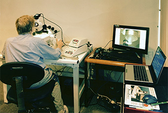
| Sample preparation for AFM studies, a crucial part of all experiments, can be substantially helpful in advanced characterization of different materials. Like all other characterization techniques, AFM has some limitations and is best applied to smooth samples with small corrugations that can be efficiently profiled with a microscopic probe having a tip apex of several nanometers in radius. Therefore, polymer samples best suited for AFM examination are prepared by hot pressing between two smooth surfaces (glass slides, pieces of Si wafer, etc) or by spin-casting the polymer solutions on different substrates. However, in many cases, researchers are particularly interested in the bulk morphology of a polymer sample. The only way to provide access of AFM probe to the sample interior is to break or cut the prepared sample. Ultramicrotomy performed with sharp diamond knifes is the most useful technique to prepare polymer samples for studies of their bulk morphology. As many polymer materials include rubbery inclusions and components, whose glass transition temperatures are well below room temperature, their sample preparations need to be performed at cryogenic temperatures. Therefore, cry-ultramicrotomy of polymer samples is often used for AFM analysis. A few weeks ago, member of NT-MDT visited one of the manufacturers of RMC cryo-ultramicrotomes (Boeckeler Instruments Inc) in Tucson, Arizona. The engineers at this company kindly helped us with a customer sample preparation, seen in the photo below. In further expansion of ultramicrotomy applications, Boeckeler Instruments recently introduced an automatic tape collecting microtome that continuously makes multiple sections of a sample and deposits them on a substrate such as polyimide tape of Si wafers. |
 Dr. Greg Becker (Boeckeler Instruments) is working with RMC cryo-ultramicrotome. A successive examination of these sections with any microscopes including AFM helps to reconstruct the three-dimension profiles of sample structure. Such tomography is in high demand for structural studies of biological objects, such as the brain, and multicomponent polymer materials. |
|
| Current Marketing Activities | ||
| Our recent developments have led to the introduction of new imaging modes such as amplitude modulation with frequency imaging (AM-FI) and frequency modulation (FM), which complement a more traditional amplitude modulation with phase imaging (AM-FI) and Hybrid mode. In exploring the capabilities of new modes, we found that in AM-FI a lowering of set-point amplitude leads to monotonic increase of the peak tip-force in resonant oscillations and the frequency contrast correlates with elastic modulus variations. A precise control of the tip-sample forces in the attractive and repulsive force regimes is offered in FM mode. | A detailed description of AFM operations in the new mode is the subject of a new Application Note: “Exploring Imaging in Oscillatory Resonance AFM Modes: Backgrounds and Applications”. This document can be found at the following link: size: A4 or Letter In another attempt to bring our innovations to the attention of the AFM community, we have upgraded the NT-MDT corporate brochure, and the new edition can be found at the following link: Scanning Probe Microscopes. NT-MDT Corporate Brochure |
|
| Summer Time! | |||||
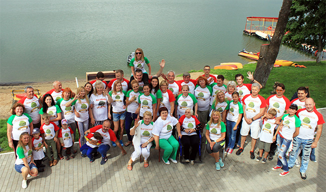
| Summer months are traditional vacation time and our employers have used it to escape from the Arizona heat and to visit more comfortable locations. Elena and Sergei Magonov, along with their daughters, spent some time in Europe and attended their family reunion that took place at a lake resort in Belarus - the country where Sergei was born. |
This event attracted over 50 people from the Magonov family who came to this gathering from 7 countries including Cyprus, the Netherlands, Chile, and the USA. This tradition that helps maintain family ties through generations made the participants very joyful and excited as seen from the picture below. | ||||
 |
|||||
| Cool AFM Images | |||||
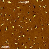
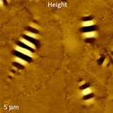
| Our gallery of attractive AFM images has been enriched by new images of bee-like structures that were recorded from various bitumen samples. Two height images, which were obtained in AM-PI mode, reveal a complex morphology of industrially important material that serves as an asphalt binder. This AFM study, which is aimed at understanding the bitumen morphology, is a part of joint efforts with Prof. Elham Fini and Daniel Oldham (NC A&T SU) who kindly provided the samples. |
|
||||
| Forthcoming Events | |||||
| On September 3, 2015, Dr. Sergei Magonov (Tempe, AZ) will participate in the NT-MDT Workshop in Basel (Switzerland), where he will present a lecture on “Expanding Atomic Force Microscopy Studies to Quantitative and High-Resolution Mapping of Local Mechanical and Electrical Properties”. | |||||


